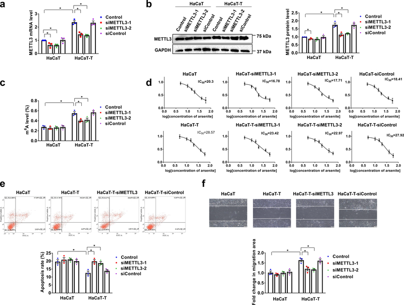
Fig. 2. m6A mediates migration and apoptosis in arsenite-transformed keratinocytes.
a, b, The mRNA (a) and protein (b) levels of METTL3 in HaCaT and HaCaT-T cells with introduction of two siMETTL3 (siMETTL3–1 and siMETTL3–2) and siControl were tested by RT-qPCR and western blotting, respectively. Representative images of western blotting (b, left). c, Total m6A levels in HaCaT and HaCaT-T cells with introduction of siMETTL3 and siControl were determined by EpiQuik m6A RNA Methylation Quantification Kit. d, Cell viability of HaCaT and HaCaT-T cells with different treatments was tested by MTT assay. e, Representative images of apoptosis tested by flow cytometry (up) and quantification of cell apoptosis in HaCaT cells and HaCaT-T cells with induction of two siMETTL3 (siMETTL3–1 and siMETTL3–2) and siControl (down). f, Representative images of cell migration examined by wound healing assay with a microscope at 400× magnification (up) and quantification of migration in HaCaT cells and HaCaT-T cells with induction of two siMETTL3 (siMETTL3–1 and siMETTL3–2) and siControl (down). For a-f, n = 3 biological replicates, error bars indicate mean ± SEM. The P-values were determined by two-tailed t-test. “*” indicates a significant difference compared to control group (P < 0.05).

