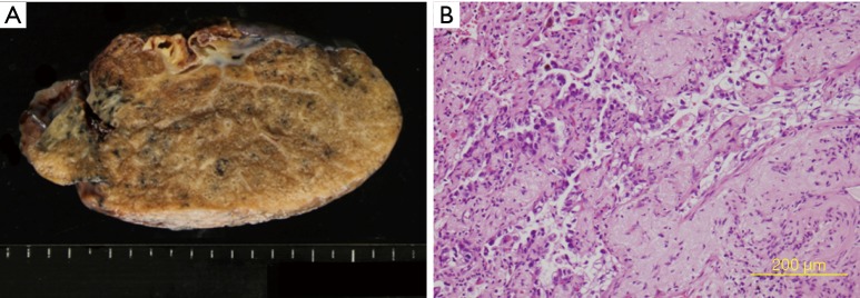
Figure 4.
Pathological findings. (A) Macroscopic findings of the resected specimen. The tumor was 13 mm × 6 mm in size, and intrapulmonary metastasis was observed (H&E stain). (B) The postoperative pathological diagnosis was invasive adenocarcinoma with N1 lymph node metastasis. Chemotherapy effectiveness was rated as EF2, ypT3(PM1)pN1cM0 stage IIIA.

