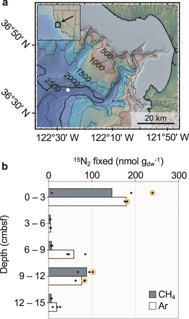
Fig. 1. Sampling location and previously measured N2 fixation in the sediment core used in this study.

a Map of sampling site at Monterey Canyon, California, USA. Sampled location marked with a white circle. Contour lines show 500 m depth intervals. b 15N2 assimilation in sediment microcosms measured using isotope ratio mass spectrometry (reproduced from Dekas et al. [12]). Microcosm headspace gas is indicated. Each circle represents a biological replicate, with bars indicating the average 15N2 assimilation. Large, yellow circles indicate the replicate bottle used for 15N-SIP and nifH analyses.
