Fig. 1.
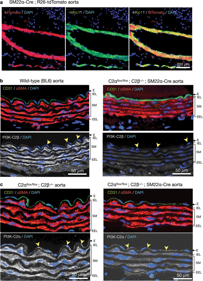
Deletion of C2α and C2β in the aortic smooth muscle layer of knockout mice. a The expression of tdTomato and immunofluorescence staining of myosin heavy chain 11 (Mhc11) in the medial smooth muscle layer of the aorta in SM22α-Cre; R26-tdTomato reporter mice. Mhc11-positive cells express tdTomato protein. b Deletion of C2β in the medial smooth muscle layer and the endothelium of the aorta in C2αflox/flox C2β−/−; SM22α-Cre mouse. The sections of the aortic wall were immunostained using anti-C2β, anti-CD31 and anti-αSMA antibodies. The medial smooth muscle layer (SM) and the endothelium (E) were completely devoid of C2β expression in C2αflox/flox C2β−/−; SM22α-Cre mouse (lower right), differently from wild type (BL6) mouse (lower left). IEL internal elastic lamina, EEL external elastic lamina. c Marked reduction of C2α expression in the SM but not the E of the aorta in C2αflox/flox C2β−/−; SM22α-Cre mouse. The sections of the aortic wall were immunostained using anti-C2α, anti-CD31 and anti-αSMA antibodies. In b and c, yellow arrowheads indicate endothelial deletion of C2β in C2αflox/flox C2β−/− mouse but not of C2α in C2αflox/flox C2β−/−; SM22α-Cre mouse, respectively. In a–c, nuclei were stained with DAPI

