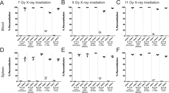
Figure 1.

Chimerism achieved with various doses of X-ray irradiation. Animals were irradiated with varying doses of X-ray irradiation and analyzed 8 weeks post reconstitution to evaluate chimerism. The reconstitution of the donor hematopoietic cells (CD45.2+; closed circles) or residual recipient hematopoietic cells (CD45.1+; open squares) is shown as a percent of total cells in the blood (A-C) or in the spleen (D-F) of animals. In general, the range of irradiation used demonstrated comparable reconstitution. This figure is representative of two independent experiments. Data represents mean +/- standard deviation with 5 mice per group.
