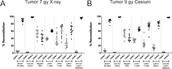
Figure 6.

Chimerism at 8 weeks post reconstitution in the tumor microenvironment of X-ray and cesium irradiated animals. Donor cell reconstitution was evaluated in animals that were irradiated with X-ray (A) or Cesium137 (B) and implanted with 4 × 10⁵ B16F10 melanoma tumor cells. Tumors were implanted and allowed to establish before dissociation and analysis on day 13 post implantation (approximately 140–220 mm3. Donor hematopoietic cells (CD45.2+; closed circles) were differentiated from recipient hematopoietic cells (CD45.1+; open squares) by flow cytometry using congenic markers. Hematopoietic cells were identified based on commonly expressed leukocyte antigens. The figure is representative of at least two independent experiments.
