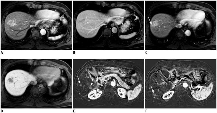
Fig. 2. MRI of 54-year-old male with HCC.
Arterial phase tumor rim enhancement, peritumoral parenchymal enhancement, and high APRI were noted. APRI level was 0.456.
In S8 dome, 3-cm sized lobulated mass showing strong enhancement on arterial phase (A) with subsequent washout on portal phase (B) is present. Of note, arterial phase enhancement is not homogeneous; enhancement is seen mostly in outer or peripheral portion of tumor while inner portion shows poor enhancement. Peritumoral parenchymal enhancement on arterial phase (arrow) (C) was noted beyond tumor margins that can be defined on hepatobiliary phase (D). E, F. Segmentectomy was performed for HCC. Seven months after surgery, subtraction of pre-contrast from arterial phase images demonstrated multiple recurrent HCCs in liver (arrows). APRI = AST/platelet ratio index, AST = aspartate aminotransferase

