Figure 1.
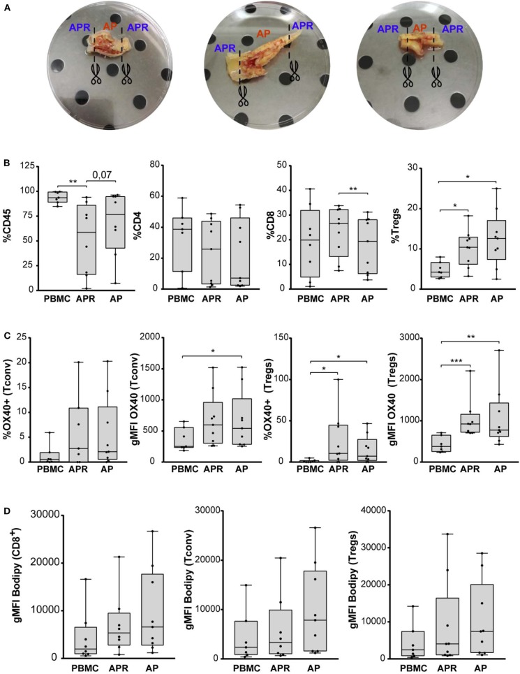
(A) Three representative carotid endarterectomy samples. The image shows sample division strategy: APR (adjacent atherosclerotic plaque region), AP (atherosclerotic plaque). (B) Frequency of CD45, CD4, CD8, and Tregs (in gated CD4+ T cells) in PBMC, APR, and AP from CEA patients (n = 9). (C) Percentage of OX40 in Tconv and Tregs and gMFI of OX40 gated on Tconv and Tregs. (D) gMFI of BODIPY in CD8, Tconv, and Tregs. In all figures, Tregs have been identified as FOXP3+ CD127low within the CD14– CD8– viability dye– CD4+ CD25+ gate. *P < 0.05, **P < 0.01, ***P < 0.001, by paired t-test, two-tailed.

