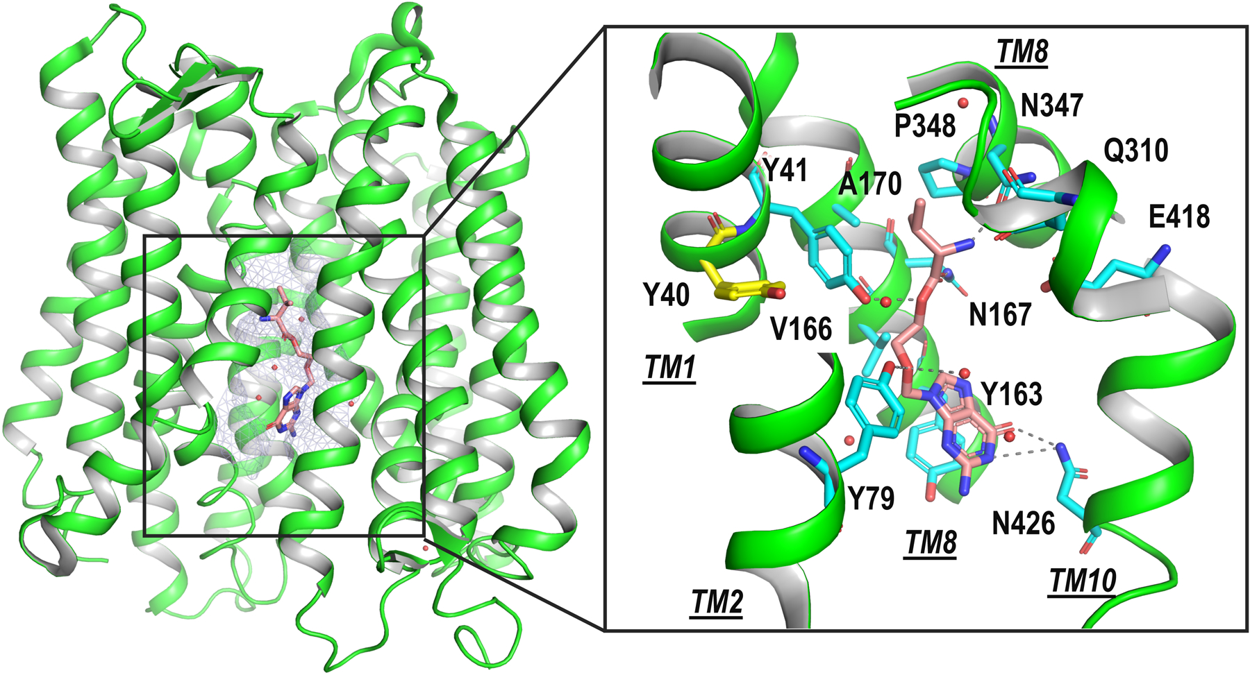
Fig. 3. Structure of an ADME transporter homolog, PepTSh.

The crystal structure of PepTSh (green cartoon) in complex with the antiviral drug valacyclovir (peach sticks) is shown. The substrate binding site is shown in light blue mesh. Inset shows the magnified view of the interaction of valacycolovir with the binding site of PepSh. The sidechain atoms of key residues in PepTSh are illustrated with cyan sticks, with Y40, in yellow. Y40 is equivalent to F28 in the human PepT1. Rare mutation in this position in African Americans (F28Y) reduces substrate uptake by PepT1. Hydrogen bonds between binding site residues and valacycolvir are displayed as dashed gray lines. Images were generated with PyMOL (https://pymol.org/2/).
