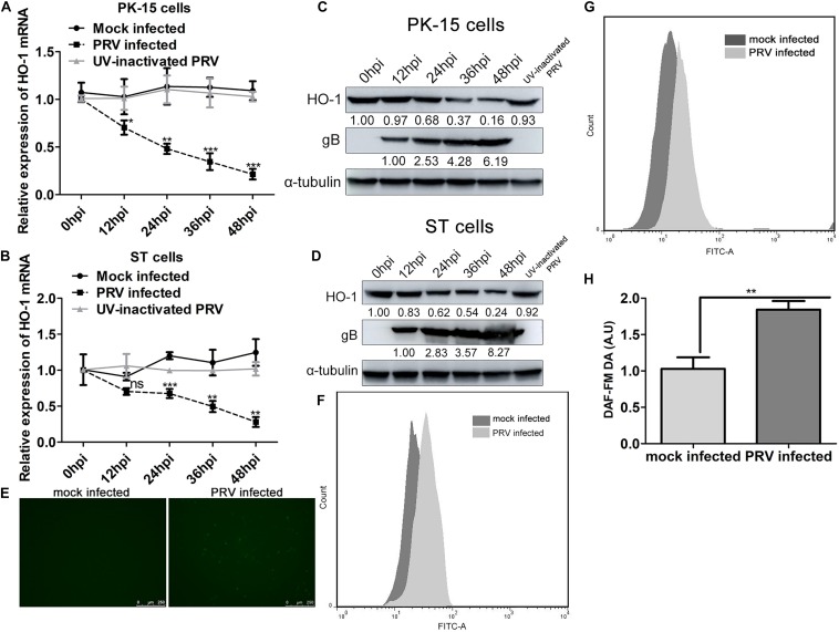
FIGURE 1.
PRV infection suppresses HO-1 expression while stimulates oxidative stress response in both PK-15 and ST cells. (A,C) PK-15 or (B,D) ST cells were infected with PRV at a MOI of 0.01 for 1 h at 37°C. Cells samples were collected at 0, 12, 24, 36, and 48 hpi to detect HO-1 and PRV gB mRNA and protein levels via RT-qPCR and western blotting, respectively. (E,F) PK-15 cells were infected with 0.01 MOI of PRV for 24 h. Then cells were incubated with DMEM supplemented with 10 μM/l of DCFH-DA at 37°C for 20 min. Cells were observed under a confocal fluorescence microscope (100x) and assessed via flow cytometry analysis. (G,H) PK-15 cells infected with 0.01 MOI of PRV for 24 h were incubated with DMEM containing 5 μM/l DAF-FM DA and incubated for 20 min at 37°C. After washing using PBS for three times, cells were subjected to flow cytometry analysis. PK-15 cells mock infected with PRV were included as a control in both experiments. Data are presented as the mean ± standard deviation of three independent experiments. *P < 0.05, **P < 0.01, ***P < 0.001. PRV, pseudorabies virus; HO-1, heme oxygenase-1; RT-qPCR, reverse transcription-quantitative PCR.

