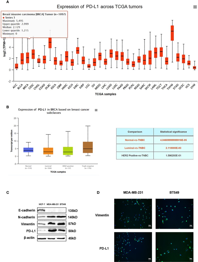
Figure 6.
PD-L1 is highly expressed in TNBC. (A) The expression of PD-L1 across TGCA tumors. (B) The expression of PD-L1 in breast cancer subtypes from TGCA database. (C) Western blot analysis of E-cadherin, N-cadherin, Vimentin, and PD-L1 in MDA-MB-231 and BT549 cells. (D) Immunofluorescence assay analysis of Vimentin and PD-L1 in MDA-MB-231 and BT549 cells.

