
Figure 3:
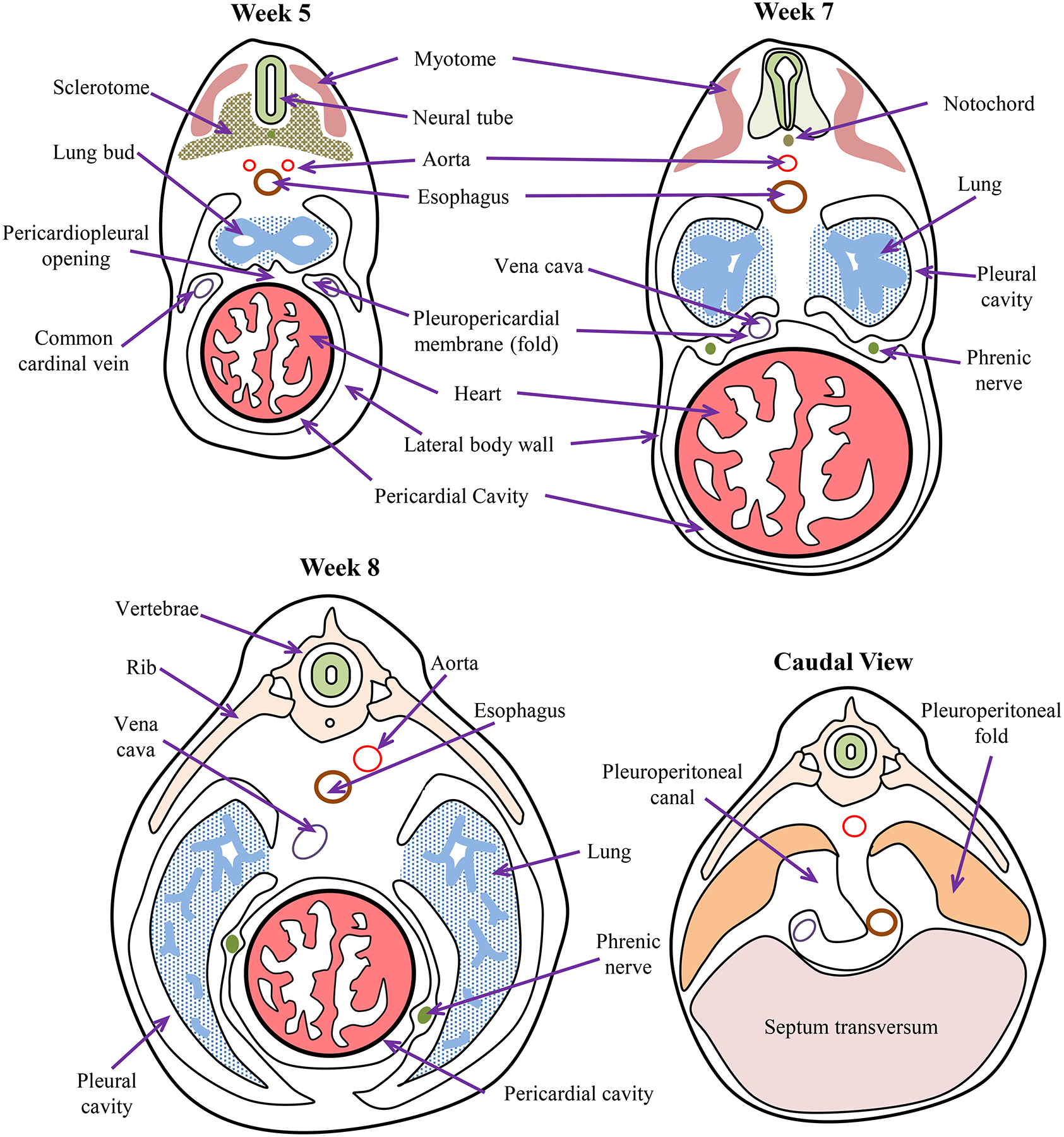
Embryonic timeline of body cavity formation in humans. By week 5, pleuroperitoneal membranes are a pair of membranes which gradually separate the pleural and peritoneal cavities, produced as the pleural cavities expand by invading the body wall. The pleuropericardial membranes separate the developing heart from the developing lung buds. Pleuropericardial membranes initially appear as small folds or ridges projecting into the primitive undivided thoracic cavity. The folds contain the common cardinal veins which drain the primitive venous system into the sinus venosus of the primitive heart. During week 6, the edges of these membranous folds have fused with the dorsal mesentery of the esophagus and with the septum transversum to separate the pleural and pericardial cavities. As the heart descends and the pleural cavities expand, the membranes are drawn out in a mesentery like fold that extends from the lateral wall. By week 7, these membranes fuse with the mesoderm ventral to the esophagus, forming a single pericardial cavity and left and right pleural cavities. During week 8, the lung buds grow into the medial walls of the nascent pleural cavities; and the pleural cavities expand around the heart into the body wall. After Pansky [496].