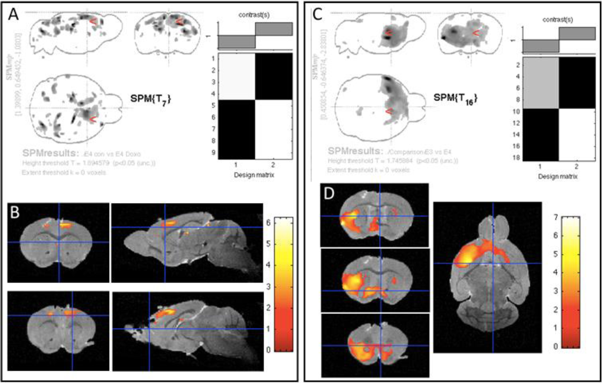
Figure 6: Long-term effects of doxorubicin on the brain structure of APOE mice are modest.

Nine months after treatment, 3-D MRI and VMB were performed on control and doxorubicin-treated APOE3 and APOE4 mice. A and C show maximum intensity projections (MIP) and the design of the matrix for the study. B and D depict color overlays of the t-test values on the co-registered template image and the location of significant clusters in this comparison. The color bar measures t test values of the statistical analyses. A & B. VBM analysis comparing the grey matter of untreated versus doxorubicin-treated APOE4 mice revealed a mild atrophy predominantly in the frontal cortex (colored areas). APOE3 mice did not show any long-term brain regional differences in response to doxorubicin (data not shown). C & D. The comparison of all (both treated and untreated) APOE3 mice versus all APOE4 mice, detected regions of grey matter that were significantly larger in APOE3 mice than in APOE4 mice, regardless of the treatment.
