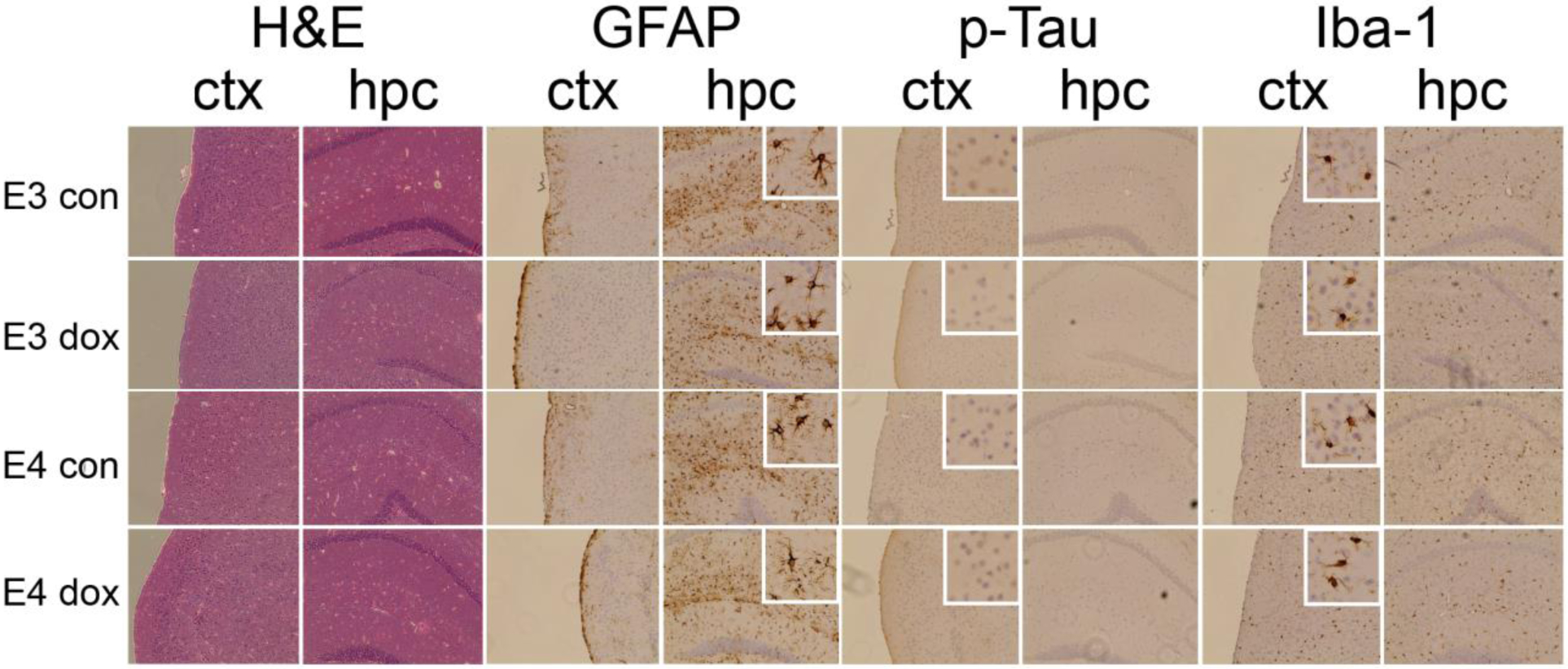
Figure 7: Immunohistochemical staining for pathogenic markers of AD.

Coronal sections across the anterior/posterior brain regions were stained from 3–5 brains of APOE3 and APOE4 mice treated with control (con) or doxorubicin (dox). Images show staining of frontal cortex (ctx) and hippocampus (hip): Hematoxylin and eosin staining (H&E), IBA-1, GFAP, phosphorylated tau. Images were taken using brightfield microscopy at 20× magnification.
