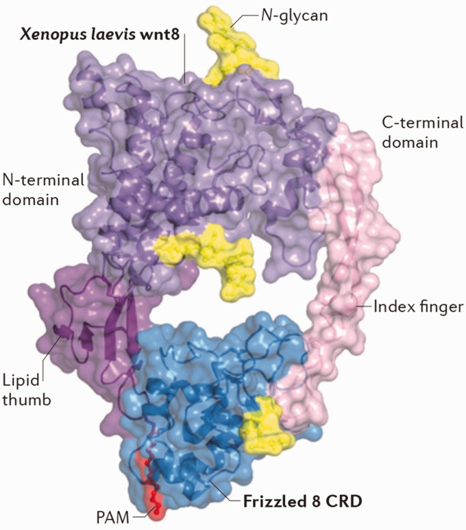
Figure 1.
Crystal structure of the Wnt–Frizzled binding complex. Overall cryo-EM structure of the interaction between X. laevis Wnt8 and the CRD of Frizzled 8 receptor, shown in a ‘face on’ presentation. Purple: the core of the X. laevis Wnt8. Deep purple: lipid thumb domain on the amino-terminal (N-terminal) of the X. laevis Wnt8. Light pink: The index finger domain on the carboxy-terminal of the X. laevis Wnt8. Red: the palmitoleic acid motif on the X. laevis Wnt8. Yellow: N-glycans motifs on the X. laevis Wnt8. Blue: The CRD of the Frizzle 8 receptor. Source: Figure cited from Niehrs.19
CRD: cysteine rich domain; PAM: palmitoleic acid motif.

