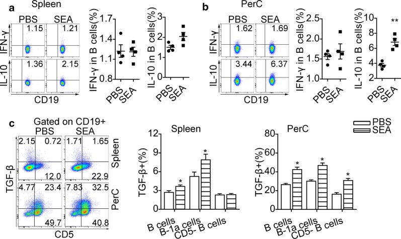
Fig. 4.
SEA-stimulated B cells acquire elevated expression of IL-10 and TGF-β in vitro. Splenic B cells and PerC washout cells from uninfected mice were stimulated with SEA for 24 h. Intracellular IL-10 and IFN-γ staining were performed after the restimulation with PMA and ionomycin in the presence of Brefeldin A. Representative flow cytometric plots and significance from statistical analyses of the percentages of IL-10+ B cells and IFN-γ+ B cells in splenic B cells (a) and PerC B cells (b) are shown. c Representative plots of CD5 and TGF-β expression on CD19+ B cells and the percentages of B cell subsets expressing TGF-β. All data are representative results of two independent experiments with at least 4 mice per group. *P < 0.05, **P < 0.01

