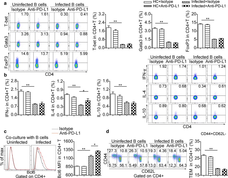
Fig. 6.
B Cells from infected mice modulate the phenotype of CD4+ T Cells in vivo. Purified splenic B cells from uninfected mice and infected mice (eight weeks) were treated with isotype antibody or anti-PD-L1 antibody respectively prior to co-culture with CD4+ T cells. a Representative dot plots of T-bet, Gata3, and FoxP3 in CD4+ T cells and significance levels from statistical analysis. b Representative flow cytometric plots of IL-10, IL-4 and IFN-γ in CD4+ T cells and significance levels from statistical analyses. c Representative flow cytometric histograms of Bcl6 expression in CD4+ T cells and significance levels from statistical analysis. The shaded histograms represent the FMO control. d Representative dot plots of CD44 versus CD62L gated on CD4+ T cells (CD44+CD62L− T effector memory, TEM) and significance levels from statistical analysis. All data are representative results of two independent experiments with at least 4 mice per group. *P < 0.05, **P < 0.01

