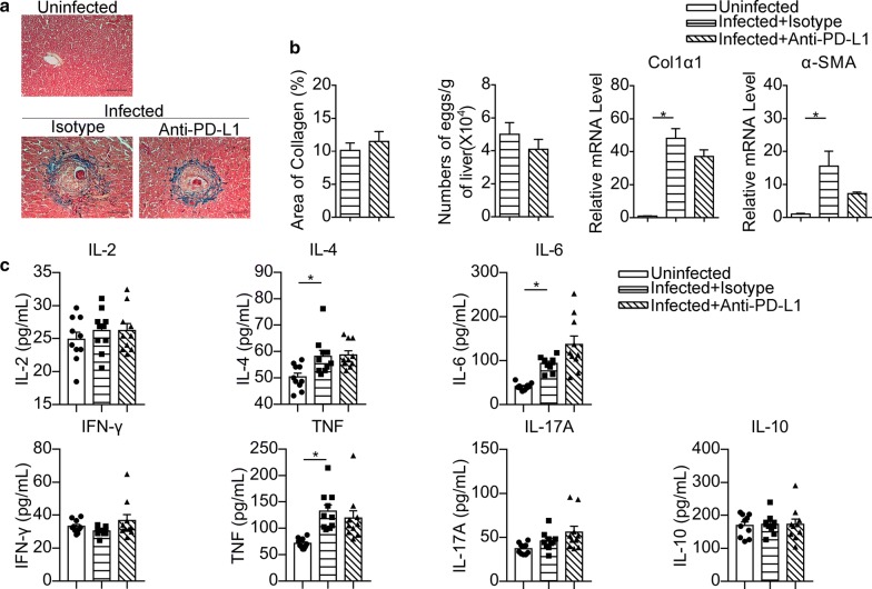
Fig. 9.
PD-L1 blocking fails to affect hepatic pathology and serum cytokines during infection. a Representative images of Masson’s trichrome staining for hepatic fibrosis analysis (original magnification of 200×). b Quantification of hepatic collagen deposition and egg burden are shown, and total RNA was extracted from livers and analyzed by RT-PCR for the expression of Col1a1 and a-SMA. In a and b, the data are representative results of two independent experiments with at least 4 mice per group. c The serum levels of IL-2, IL-4, IL-6, IL-10, IL-17A, TNF and IFN-γ were assayed by CBA. The data represent the cumulative results of two independent experiments. *P < 0.05. Scale-bars: a, 100 μm

