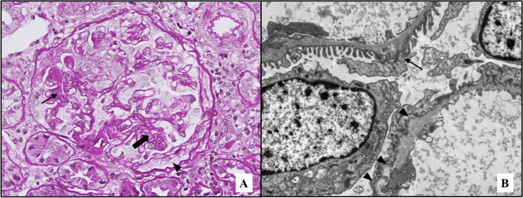
Figure 1: Representative images of renal biopsy.
(A) Glomerulus showing foci of segmental sclerosis (arrow), as well as some capillary collapse (thick arrow). In addition, a focus of prominent epithelial cells (pseudocrescent) is present (arrowhead). PAS stain, original magnification 40X. (B) Ultrastructural examination revealed only focal foot process effacement, together with diffuse thinning of the glomerular basement membranes (mean 224 nm, SD 47 nm), as seen in the upper capillary wall (arrow). Rare foci of thickening, together with splitting and scalloping of the outer contour of the lamina densa were identified (arrowheads), as seen in the lower capillary wall. Original magnification 8,000X.

