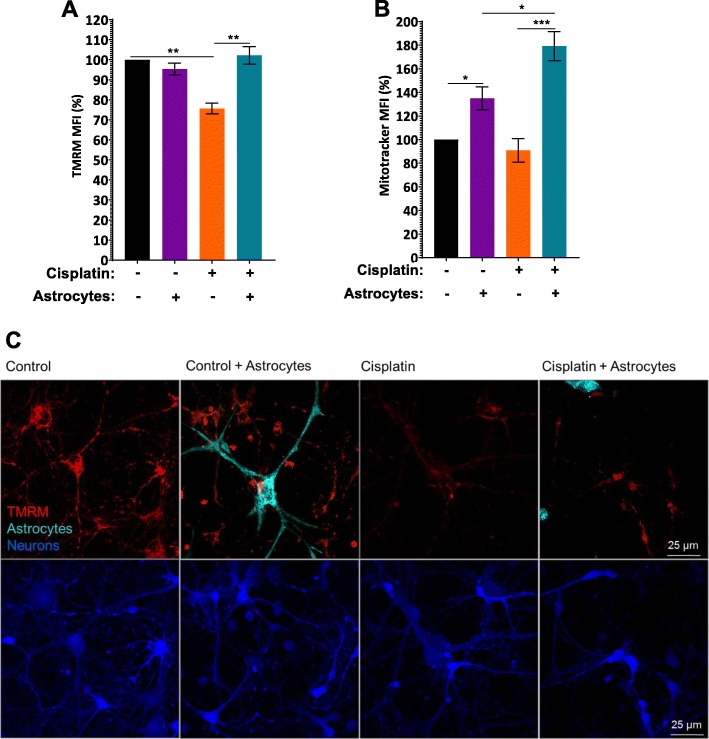
Fig. 2.
Astrocytes improve mitochondrial membrane potential of neurons damaged by cisplatin. After treating neurons and astrocytes separately with and without cisplatin for 24 h, neurons were labeled with Celltracker Green (CTG) and co-cultured with astrocytes for 17 h. Cells were labeled with TMRM (a) to assess mitochondrial membrane potential or with mitotracker (b) to quantify mitochondrial content by FACS analysis. Fluorescence intensity was normalized to fluorescence intensity in control CTG-positive neurons for each experiment and data represents the mean ± SEM of 3 independent experiments performed in duplicate. Two-way ANOVA TMRM: cisplatin x astrocyte interaction: *p < 0.05; Tukey’s post-hoc test: **p < 0.01. Mitotracker: cisplatin x astrocyte interaction: *p < 0.05; Tukey post-test: *p < 0.05, ***p < 0.001. c. Representative confocal images of the TMRM signal (red) with neurons labeled with CTB and astrocytes with cell tracker deep red (Teal). Scale bar: 25 μm

