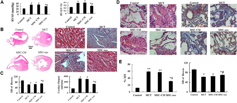
Fig. 2.
Effect of MSC-exo MCT-induced pulmonary artery pressure, vascular remodeling and right ventricle hypertrophy. a Comparative analysis of RVSP and RV/(LV + S) ratios (b) Morphology and histology of the heart were observed by hematoxylin and eosin (HE) and Masson’s staining. c Comparative analysis of fibrosis and cardiac output. d Pulmonary arterioles wall thickness and fibrosis were observed by HE and Masson’s staining. e Comparative analysis of the percent of muscular artery (MT%) and optical density (OD). n = 5 rats per group; P < 0.05, t-test; the data are present as mean ± SD; *MCT or *MSC-CM vs. control; #MSC-exo vs. MCT group; scale bar = 100 μm

