Fig. 1.
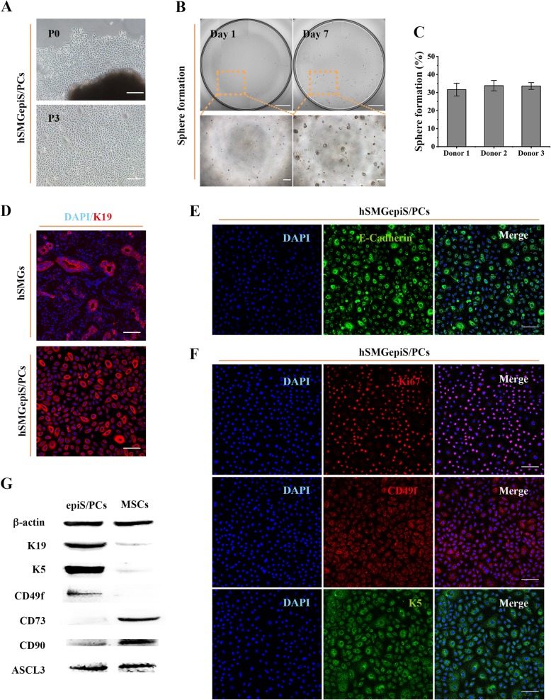
Isolation, identification, and characterization of hSMGepiS/PCs. a Images of primary cells isolated and passaged in 2D culture. Scale bar = 200 μm. b Representative images of single cells 3D-cultured and spheres formed. Scale bar = 1 mm (above) and 200 μm (below). c Quantification of sphere formation of hSMGepiS/PCs from different donors at day 7 after 3D culture. Error bars, SD, n = 6. d Immunofluorescence of K19 in cells and hSMGs to locate the isolated cells. Scale bar = 100 μm. e Positive expression of an epithelial marker, E-cadherin, in isolated cells. Scale bar = 100 μm. f Immunofluorescence showing that isolated cells express the proliferation marker, Ki67, and the epithelial stem/progenitor cell markers, K5 and CD49f. Scale bar = 100 μm. g Protein levels of the indicated markers in hSMGepiS/PCs and hSMGMCs by western blotting

