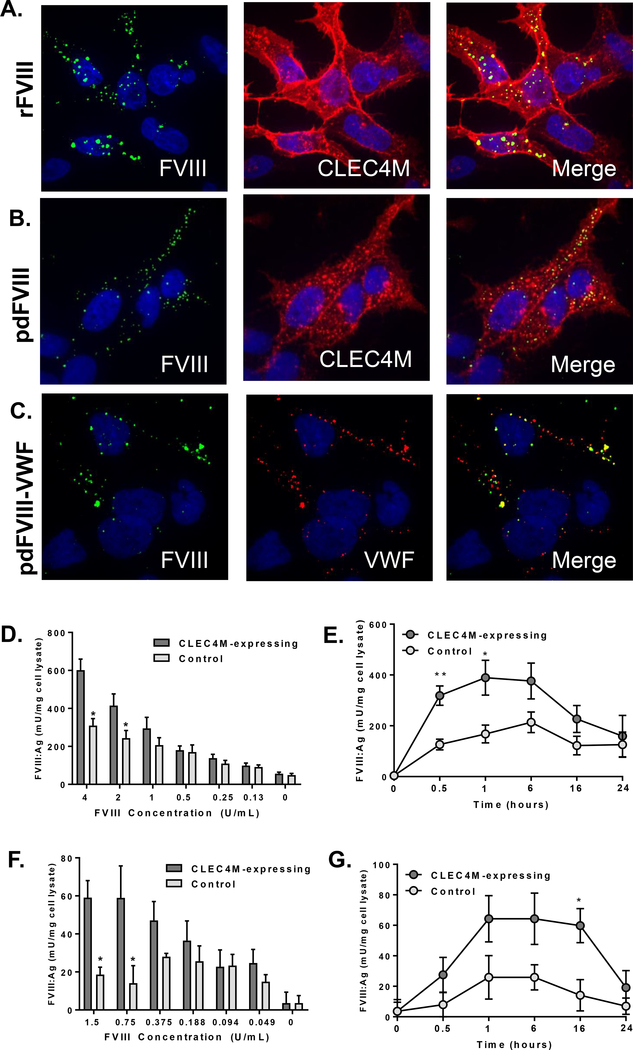
Figure 1. Internalization of FVIII by CLEC4M-expressing HEK 293 cells.
CLEC4M-expressing cells were exposed to recombinant (r) or plasma-derived (pd) FVIII and internalization was measured using immunofluorescence or ELISA. (A) Internalization of rFVIII, (B) pdFVIII by CLEC4M expressing cells using immunofluoresence (2 U/mL FVIII, 1 hour incubation); CLEC4M (red), FVIII (green), DAPI (blue), colocalization (yellow). (C) Internalization of VWF-FVIII complex by CLEC4M-expressing cells; VWF (red), FVIII (green), DAPI (blue), colocalization (yellow) (2 U/mL VWF, 0.83 U/mL FVIII, 1 hour incubation). Images are representative of 5–6 independent experiments. Quantification of rFVIII internalization by CLEC4M-expressing cells by FVIII:Ag ELISA; dose response (1 hour) (D), time course (2 U/mL FVIII) (E). Quantification of pd-VWF-FVIII internalization by CLEC4M-expressing cells by FVIII:Ag ELISA; dose response (1 hour) (F), time course (1.5 U/mL FVIII) (G). For all ELISA conditions, n=3–5 independent experiments. ± SEM, *p<0.05, **p<0.001.

