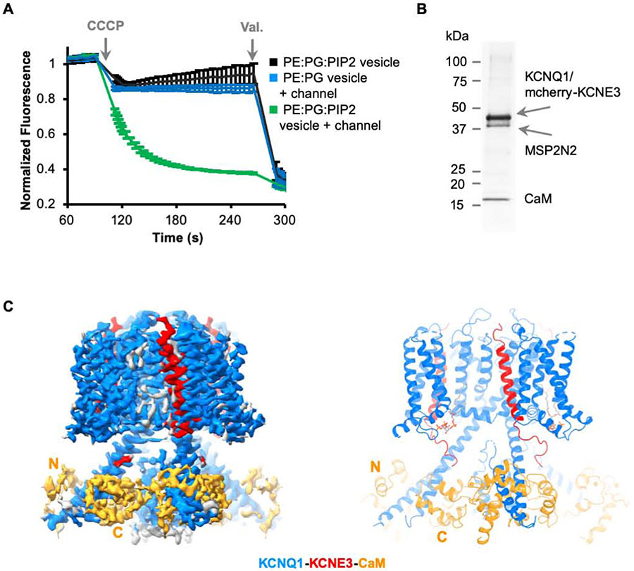
Figure 3. Structure of hKCNQ1EM-KCNE3-CaM complex in the presence of PIP2.
(A) Liposome based flux assay. Recordings (n=3) from PE:PG:PIP2 alone, PE:PG with hKCNQ1EM-KCNE3-CaM and PE:PG:PIP2 with hKCNQ1EM-KCNE3-CaM vesicles are plotted in black, blue and green traces.
(B) SDS-PAGE showing the reconstitution of hKCNQ1EM-KCNE3-CaM complex in nanodiscs containing PIP2 using MSP2N2 as the scaffold protein.
(C) Cryo-EM map and structure model of hKCNQ1EM-KCNE3-CaM in the presence of PIP2. KCNQ1, KCNE3 and CaM are colored in blue, red and orange, respectively.

