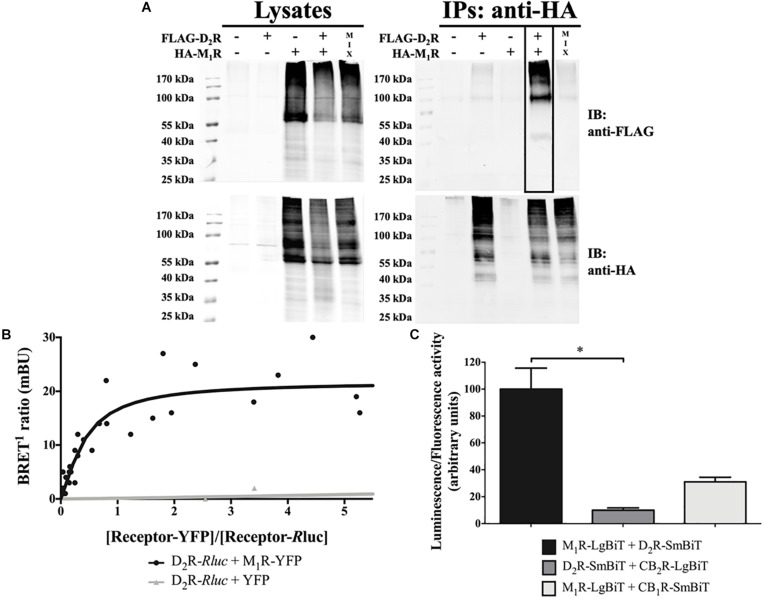
FIGURE 1.
D2R–M1R interaction in transiently transfected HEK293T cells. (A) Co-immunoprecipitation. HEK293T cells were harvested and lysed 48 h after transfection. The lysates were used for immunoblotting (IB) with anti-FLAG and anti-HA antibodies to demonstrate D2R and M1R expression, respectively (left panels). The rest of the samples (IPs) was subjected to immunoprecipitation with a mouse anti-HA antibody. The co-immunoprecipitate was confirmed via the detection of FLAG-D2R upon IB with rabbit anti-FLAG and rabbit anti-HA antibodies (right panel; boxed lane). Data shown are representative of three independent experiments. (B) BRET1 saturation curve. The BRET1 signal in HEK293T cells co-expressing a constant amount of D2R-Rluc and increasing amounts of M1R-YFP (n = 5) or YFP (n = 3) constructs was measured 48 h posttransfection. The BRET1 saturation curve is derived from all independent experiments. (C) NanoBiT® complementation assay. The SmBiT and LgBiT parts of the NanoLuciferase fragments were fused to the C-terminus of the indicated receptor. The constructs were overexpressed via transient transfection in HEK293T cells. Results are presented as mean ± SD (n = 3). Statistical significance was tested using the non-parametric ANOVA by ranks of Kruskal–Wallis followed by the Dunn multiple-comparisons post hoc test, *p ≤ 0.05.

