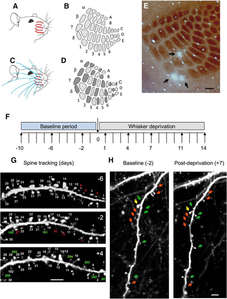
Figure 1.
Whisker deprivation patterns and spine tracking. A, Unilateral AWD, which produces (B) uniform deprivation of all barrels in the cortex. C, Unilateral CWD produces (D), a chessboard pattern of active and deprived barrels whereby every barrel deprived of its principal whisker (light gray) is surrounded by four barrels that have their principal whisker intact (dark gray) and vice versa. E, Photo-lesions are made in layer 4 of the barrel cortex on the last day of imaging (black arrows), to coregister the ROIs within which spines are imaged with their corresponding home barrels. F, Imaging time points relative to deprivation on time point 0 were −10, −6, −2, 1, 4, 7, 11, and 14 d. In some cases, 12 and 24 h time points were taken. G, Spines are tracked over a period of days, shown here for 6 d before deprivation (−6), 2 d before deprivation (−2), and 4 d after deprivation (4). Spine number 17 is branched: such cases were counted as one spine. Some spines are eliminated from one time point to the next (red numbering); others are formed anew (green numbering). H, Examples of eliminated (red arrows) and newly formed or enlarged spines (green arrows) shown for a dendrite imaged at 2 d before and 7 d after deprivation. Yellow arrow indicates a spine where the spine head shrinks over this period. Scale bars: E, 150 μm; G, H, 5 μm.

