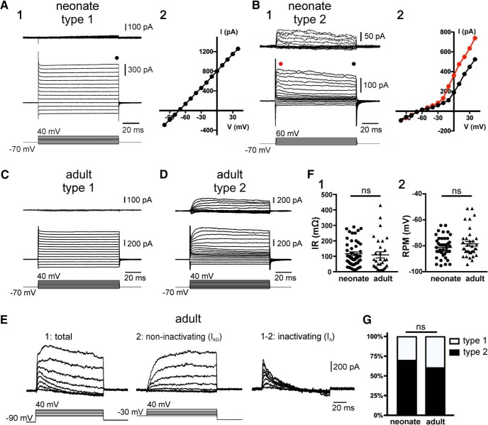
Figure 1.
Electrophysiological properties of ependymal cells in the neonatal and adult spinal cord of mice. A, Currents induced by a series of voltage steps applied to an ependymal cell on the lateral domain of the CC (A1) of a neonate mouse. The leak-subtracted currents (A1, top) and the I–V plot (A2) show that the response to voltage steps was completely passive (Type 1 cell). B, A subpopulation (Type 2) of ependymal cells in the neonatal spinal cord displayed voltage-gated outward currents (B1). The I–V plot in Type 2 cells is nonlinear (B2). The current plots at the beginning (red dots) and the end (black dots) suggest that voltage-gated outward currents have a component with time- and voltage-dependent inactivation. C, D, The electrophysiological phenotypes of ependymal cells in the adult spinal cord (P > 90) were similar to their neonatal counterpart with Type 1 passive cells (C) and Type 2 cells (D). E, Current responses to voltage steps applied from a holding potential of −90 mV (E1) and −30 mV (E2) in an ependymal cell of the adult spinal cord. Notice the slow kinetics and lack or inactivation of the outward currents from a holding potential of −30 mV typical of a delayed rectifier (IKD). Subtraction of currents at −90 and −30 mV reveals a fast inactivating component characteristic of A-type (IA) currents. F, No statistically significant differences were found for the IRs (F1, p = 0.29) and RMPs (F2, p = 0.3) between neonate and adult ependymal cells. G, Ratio of Type 1 and Type 2 cells in neonate and adult ependyma. ns, non significant.

