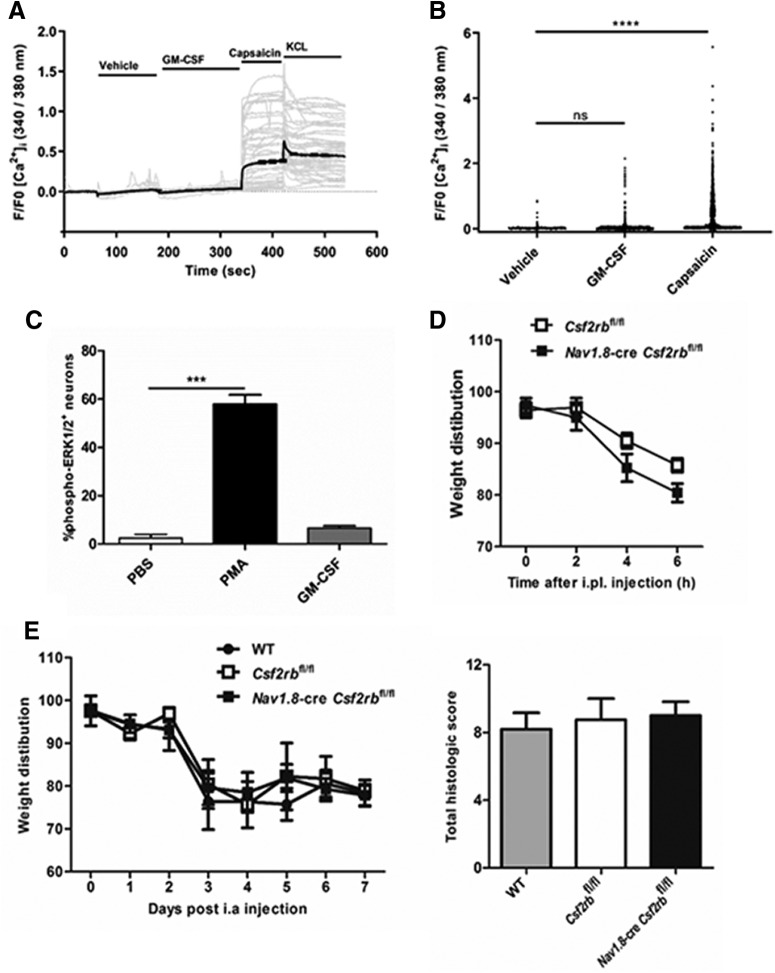
Figure 2.
GM-CSF does not directly activate neurons in vitro and in vivo. A, B, Time course and peak Ca2+ responses in mixed DRG cultures in response to vehicle, GM-CSF (200 ng/ml), capsaicin (0.5 μm), and KCl (50 mm; only A), respectively. A, Gray lines, Individual traces from 50 random cells; black lines, mean response; (B) n = 1767 neurons (pooled data from two independent experiments). C, Percentage of DRG neurons positive for phospho-ERK1/2 following stimulation with PBS, PMA, or GM-CSF (200 ng/ml) for 15 min. Three independent experiments were performed. D, E, Pain development (incapacitance meter, ratio of weight bearing on injected relative to non-injected knee/hindpaw, a value <100 indicates pain) was measured following (D) intra-planatar (i.pl.) injection of GM-CSF (20 ng) in Csf2rbfl/fl and Nav1.8-cre Csf2rbfl/fl mice (n = 5–8 mice/group); and (E) mBSA/GM-CSF arthritis [mBSA intra-articular (i.a.) (Day 0); GM-CSF or saline subcutaneously (Days 0–2)] induction in WT, Csf2rbfl/fl, and Nav1.8-cre Csf2rbfl/fl mice (n = 4–7 mice/group). Arthritis (histology, Day 7) was also assessed in E. C–E, Data are expressed as mean ± SEM. For B and C, a one-way ANOVA was used. ***p < 0.001, ****p < 0.0001.

