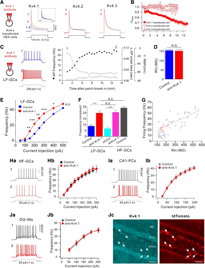
Figure 4.
Kv4.1 inhibition increases firing frequency selectively in LF-GCs. A, The specificity of Kv4.1 antibody for the Kv4.1-mediated current was determined in HEK293 cells expressing Kv4.1, Kv4.2 or Kv4.3. Outward currents evoked by depolarizing pulse to +40 mV for 1 s in Kv4.1, Kv4.2 or Kv4.3-expressing HEK293 cells measured just after patch break-in (1, black) and >12 min after intracellular application of the Kv4.1 antibody (2, red). Inset, K+ currents measured before (black) and after (red) inhibition of Kv4.1 and subtracted current (blue) in the Kv4.1-expressing cell were normalized to the peak amplitude of current. B, Summary graph of inhibition of I/Imax by Kv4.1 antibody in HEK293 cells expressing Kv4.1 (closed circle), Kv4.2 (open circle), or Kv4.3 (open triangles). Values indicate mean ± SEM. Error bars indicate SEM. C–H, Effects of Kv4.1 antibody on intrinsic excitability in LF-GCs (C–G) and HF-GCs (H). C, Example traces (left) of APs at the time points (1, 2) indicated on the right graph. Right, Graphs showing the time course of the alterations in RMP (open squares), firing frequency (filled circles) and first AP onset time to 1 s current injection (open circles) during Kv4.1 antibody perfusion in LF-GCs. D, Mean values for Rin (n = 6) measured within 3 min after patch break-in and after the changes in firing frequency induced by Kv4.1 antibody reached steady state (12 min). E, The F–I curve measured from LF-GCs in the presence of Kv4.1 antibody in the pipette solution (red closed circles, n = 10). The same data shown in Figure 3E (blue closed circles, LF-GCs) are superimposed to compare with the F–I curve obtained in the presence of Kv4.1 antibody. Significance levels for the difference between LF-GCs and anti-Kv4.1 were indicated. P values at 150, 200, 250, 300, 400, and 500 pA were 0.0024, <0.0001, 0.0048, <0.0001, 0.0035, and 0.189, respectively. F, I–O gain between 100 and 200 pA input currents obtained from each condition. Increases in I–O gain by Kv4.1 inhibition were significant (p < 0.001) *p < 0.05, **p < 0.01, ***p < 0.001, N.S. (not significant) p > 0.05 by Student's t-test. G, Firing frequency of GCs under control condition (blue circles for LF-GCs; black circles for HF-GCs) and Kv4.1-inhibited conditions using shKv4.1 or Kv4.1 antibody (red circles) was plotted against Rin. The firing frequency of HF-GCs (black circles) and Kv4.1-depleted LF-GCs (red circles) showed a linear relationship with Rin, whereas the firing frequency of LF-GCs (blue circles) is lower. H, The Kv4.1 antibody had no effects on intrinsic excitability in HF-GCs. Ha, Representative traces of AP trains obtained from HF-GCs recorded right after patch break-in (1, black) and 15 min after intracellular application of Kv4.1 antibody (2, red). Hb, Inhibition of Kv4.1 had no effect on the firing frequency of HF-GCs (n = 4). Ia, Representative traces from CA1-PCs recorded right after patch break-in (1, gray) and 15 min after intracellular application of Kv4.1 antibody (2, red). Ib, Firing frequency was plotted against the injecting currents (1 s) with or without Kv4.1 antibody in the pipette for CA1-PCs (n = 7). Ja, Representative traces of AP trains in DG interneurons from Vgat-ires-Cre mice infected with AAV-DIO-mCherry recorded at 3 (1, black trace) and 13 (2, red trace) min after patch break-in. Jb, Relationships between the firing frequency and the current injected were not changed by Kv4.1 antibody. Jc, Hippocampal section from Vgat-tdTomato mice was coimmunostained with Kv4.1 and tdTomato antibodies. Arrows indicates Vgat-positive interneurons expressing tdTomato. Scale bar, 50 μm.

