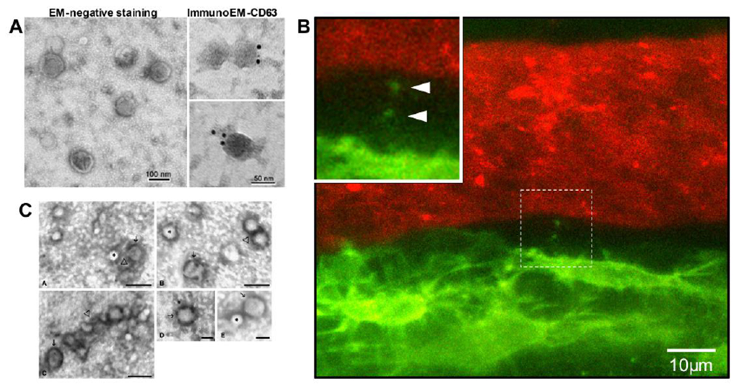
Figure 2:

A) Morphology and structure of exosomes obtained from Ocy454 osteocyte-like cells by electron microscopy analysis. The exosomes were stained with uranyl acetate (EM-negative staining) or labeled with 10-nm immunogold using antibody against the exosomal membrane marker CD63 and stained with uranyl acetate (ImmunoEM-CD63). Exosomes were visualized using a Hitachi H7000 electron microscope (reproduced from Qin et al, 2017[13], with permission from the American Society for Biochemistry and Molecular Biology). B) Still frame from intravital confocal imaging in calvarial bone of a Dmp1-mGFP transgenic mouse. Osteocytes are GFP-positive (green) and the vascular lumen is labeled with Texas Red-conjugated high MW dextran. Note an osteocyte that is releasing small vesicle-like particles adjacent to the lumen of the vascular channel (also see enlarged inset, arrowheads). C) TEM images of microvesicles released from C2C12 cells. The microvesicles were isolated from culture supernatants by 110,000 x g centrifugation, then fixed, negatively stained, and observed by TEM. The images show small vesicles of 50–80 nm in diameter. Some of these microvesicles apparently contain a diffuse electron-dense material (→) while others appear completely electron transparent (*). A tendency to aggregate (Δ) occasionally appears. Images A, B and C, bar = 100 nm; Image D and E bar = 50nm (Reproduced from Guescini et al, 2010[48] with permission from Elsevier).
