
Figure 2.
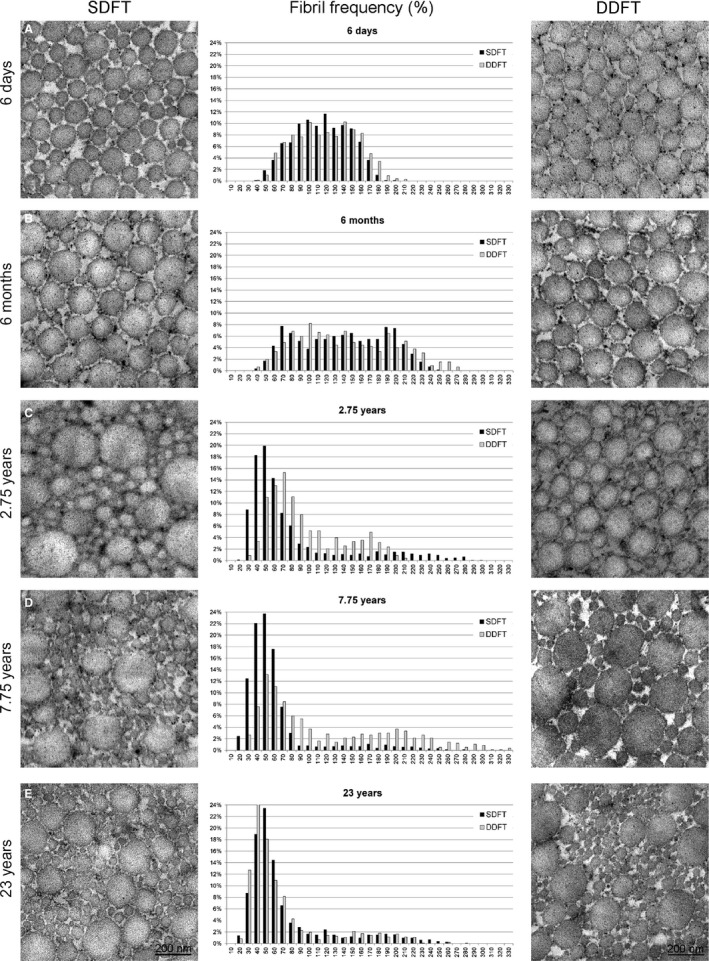
Representative TEM pictures and respective histograms showing collagen fibril distribution changes for the SDFT and DDFT with age. Whereas a Gaussian‐like distribution of fibril diameters was observed in tendons of foals up to 6 months of age, a marked shift towards much thinner fibrils was seen in tendons of horses 2.75 years of age or older.