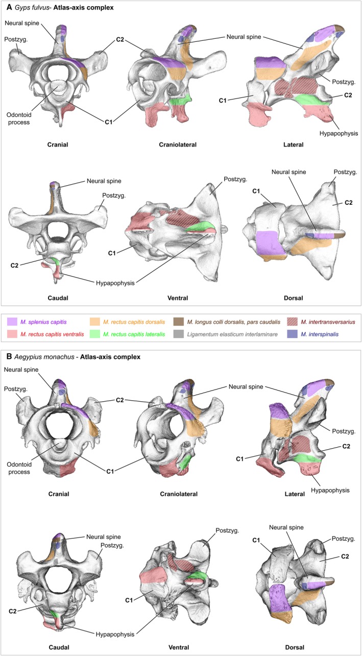
Figure 1.

Cervical vertebrae – atlas−axis complex. The first and second cervical vertebra of (A) Gyps fulvus, (B) Aegypius monachus. The two vertebrae were scanned in situ with surrounding soft‐tissue. The 3D models were reconstructed from the micro‐computer tomography (μCT) scans using the software Avizo (Version 6.3, Visualization Science Group) and the software MeshLab (ISTI‐CNR, Pisa). The areas of origin and insertion of the neck muscles are shown only for the left side of the bones. The muscles are labeled in color and the bone structures are labeled in black
