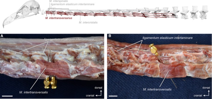
Figure 13.

Intervertebral system – lateral muscles. Top: schematic diagram (based on Gyps fulvus) visualizing the muscle topology (the relevant muscles are highlighted in color). The Mm. intertransversarii in lateral view (A) Gyps fulvus and (B) Aegypius monachus. Only one muscle belly is colored. The other intervertebral muscles have largely been removed to reveal the midline interspinous ligament. The M. intertransversarius originates laterally on the cranial part of one vertebra and inserts laterally onto the caudal part of the precedent vertebra. The Mm. intertransversarii are present between all cervical vertebrae starting from CV2 in both vultures. Scale bar: 1 cm
