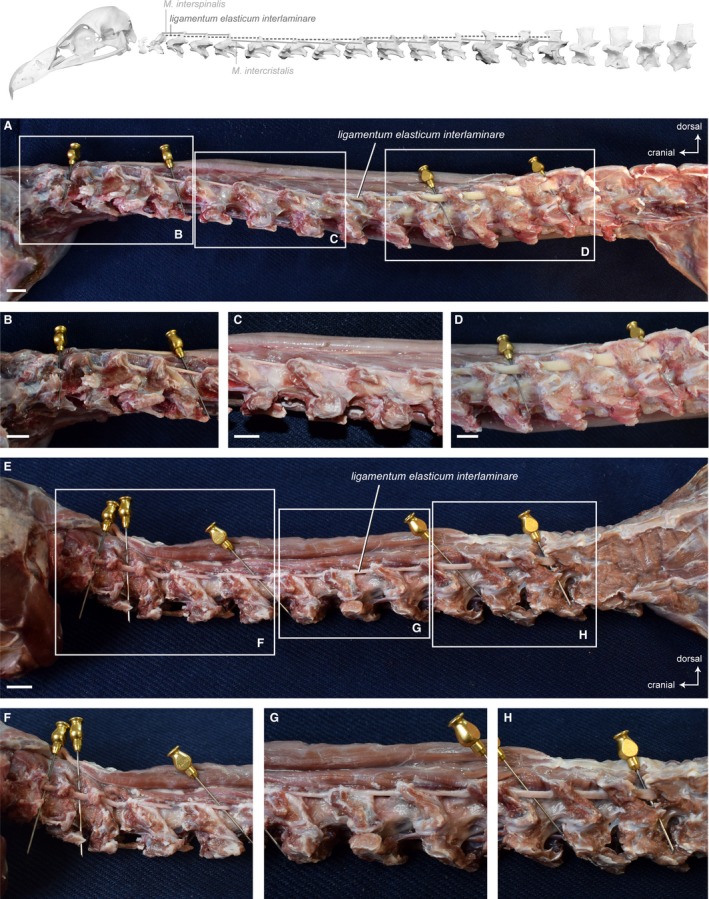
Figure 14.

Ligaments. Top: schematic diagram (based on Gyps fulvus) visualizing the muscle topology (the ligament is represented as dashed lines). The ligamentum elasticum interlaminare along the entire neck in lateral view (A) Gyps fulvus and (E) Aegypius monachus. Note the different thickness/strength of the interspinal tendons. Detailed view of the tendon in (B, F) the caudal region, (C, G) the intermediate region, (D, H) the cranial region of the neck. Scale bar: 1 cm
