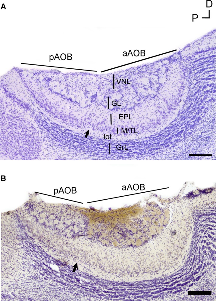
Figure 1.

Accessory olfactory bulb (AOB) anatomy of Octodon lunatus. (a) Microphotograph of a Nissl‐stained section through the medial portion of the AOB. All layers of the AOB appear readily identifiable. Note the glial indentation splitting the anterior (aAOB) and posterior (pAOB) subdomains (black arrow). (b) Anti‐Gαi2‐immunostained, Nissl‐counterstained section. The stain (brown) is exclusively restricted to the aAOB portion. EPL, external plexiform layer; GL, glomerular layer; GrL, granular cells layer; lot, lateral olfactory tract; M/TL, mitral/tufted cells layer; VNL, vomeronasal nerve layer. Scale bar: 250 μm.
