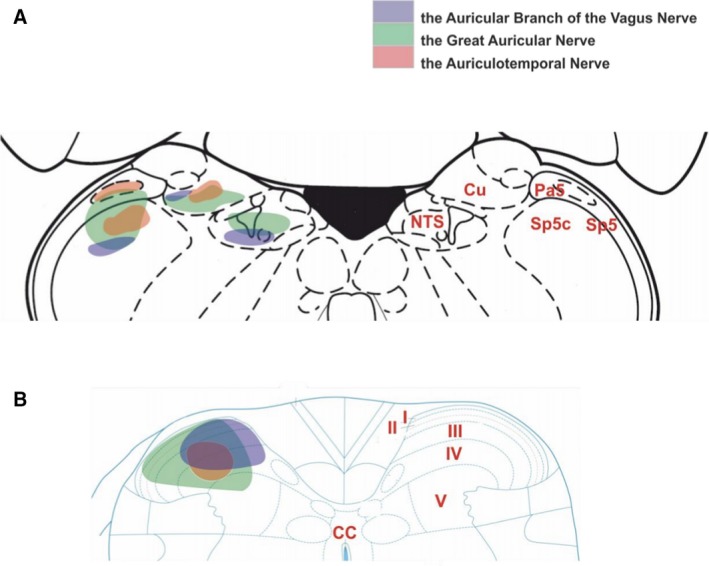
Figure 3.

A schematic diagram of external auricle afferent projections to the brainstem and upper cervical spinal cord. (A) In the brainstem, the greater auricular nerve (green) projects into the trigeminal tract, cuneate nucleus and a little in the nucleus of the solitary tract. The auriculotemporal nerve (red) terminates in the trigeminal tract, caudal trigeminal nucleus and cuneate nucleus. The ABVN (blue) is projected into the nucleus of the solitary tract, cuneate nucleus, and caudal trigeminal nucleus. (B) Projection of the greater auricular nerve into upper cervical cord has wide coverage from laminae I to laminae V, and smaller coverage by the auriculotemporal nerve concentrated in the laminae III–IV. The ABVN afferents terminate in the laminae I‐IV. The level central nervous axis was omitted for clarity. Permissions: We wish to thank Prof. Jim Deuchars (University of Leeds) and Dr Mohd Kaisan Bin Mahadi for providing this figure. No formal permissions required.
