. 2020 Feb 5;9:e51247. doi: 10.7554/eLife.51247

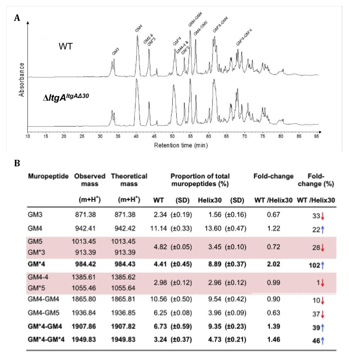
Figure 4. Muropeptide composition of PG isolated from wild-type and ΔltgAltgAΔ30.

(a) The chromatogram represents purified PG digested by the muramidse, mutanolysin and the resulting muropeptides were reduced, and analyzed by LC/MS. The results were reproducible over four biological replicates. Peak identifications correspond to (b). Quantitation and analysis of muropeptides identified by mass spectrometry. * indicates O-acetylated MurNAc. Acetylated GM*4 is highlighted in bold. Multiple muropeptides coeluted as a single peak are shaded in pink. Red arrows indicate a decrease and blue arrows an increase in muropeptide abundance. The table displays the observed and theoretical masses and the proportion of total muropeptides.

Figure 4—source data 1. Table associated with the muropeptide composition of PG isolated from wild-type, ΔltgA, ΔltgAltgA.
Figure 4—source data 2. Raw data associated with themuropeptide composition of PG isolated from wild-type and ΔltgAltgAΔ30.
elife-51247-fig4-data2.xlsx (123.4KB, xlsx)

Figure 4.

Figure 4—figure supplement 1. Muropeptide composition of PG isolated from wild-type, ΔltgA, ΔltgAltgA.
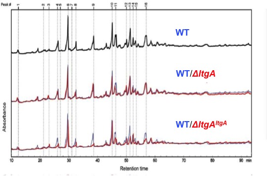
Figure 4—figure supplement 1.

The purified PG was digested by muramidase mutanolysin, and the resulting muropeptides were reduced and then analyzed by LC/MS. The results were reproducible over four biological replicates. The wild-type chromatogram (blue) is overlaid on mutant chromatograms (red). Peak numbers correspond to Supplementary file 1.
Figure 4—figure supplement 2. The expression of Ape1.
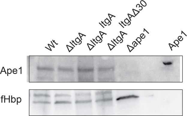
Figure 4—figure supplement 2.

The levels of Ape1 were assessed using Immunoblot and probing with anti-Ape1 antibody of the wild-type, ΔltgA, ΔltgAltgA, and ΔltgAltgAΔ30 strains. The expression of the outer membrane protein fHbp was used as an loading control. The Δape1 strain and purified recombinantly expressed Ape1 (2 ng) were used as a control to show specificity of anti-Ape1 antibody.