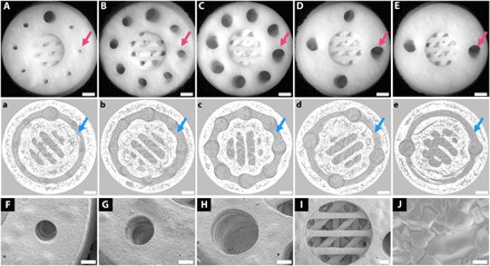
Fig. 2. 3D printing of Haversian bone–mimicking bioceramic scaffolds with cortical bone and cancellous bone structure.

Cortical bone structure contained Haversian canals and Volkmann canals. (A to E) Optical microscope images exhibited different diameters (D) and numbers (N) of Haversian canal indicated by magenta arrows (A) N = 8, D = 0.8 mm; (B) N = 8, D = 1.2 mm; (C) N = 8, D = 1.6 mm; (D) N = 4, D = 1.6 mm; and (E) N = 2, D = 1.6 mm. Scale bars, 1 mm. (a to e) Micro–computed tomography (CT) images show Volkmann canals (blue arrows) connecting Haversian canals in the interior of scaffolds. Scale bars, 1 mm. (F to J) SEM images presented the microstructure of the scaffolds. Haversian canal on the periphery of scaffolds with different diameters of (F) 0.8 mm, (G) 1.2 mm, and (H) 1.6 mm. Scale bars, 400 μm. (I) Cancellous bone structure in the center of the scaffolds. Scale bar, 400 μm. (J) Microstructure at the surface showed a well-sintered scaffold. Scale bar, 6 μm.
