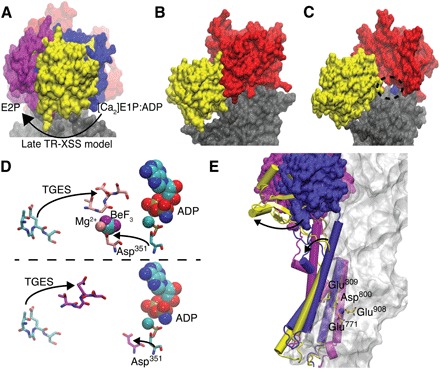
Fig. 5. Real-time dynamics compared with known structural rearrangements.

(A) Structural differences in the A domain between the late TR-XSS model (yellow), the [Ca2]E1:ADP (PDB ID: 3BA6; blue), and E2P (PDB ID: 3B9B; magenta) states. The N domain is colored red. (B) The [Ca2]E1:ADP state crystal structure (PDB ID: 3BA6) displays a closed A-N interface, while in the late-state TR-XSS model, in (C), the A domain (yellow) has moved relative the N domain (red) to expose the ADP-binding pocket (encircled). The truncated rest of the ATPase is shown in gray. (D) TGES motif and Asp351 dynamics in [Ca2]E1:ADP (PDB ID: 3BA6; cyan) and E2P (PDB ID: 3B9B; pink) (top), and [Ca2]E1:ADP (PDB ID: 3BA6; cyan) and late TR-XSS state (magenta) (bottom). (E) TM1–TM3 helices and A domains from [Ca2]E1:ADP (PDB ID: 3BA6), E2P (PDBID: 3B9B), and late state with color coding as in (A) and ion-binding residues from the late state. The truncated rest of the protein is shown as a transparent surface.
