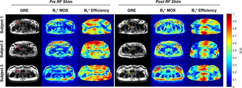
Figure 1.
The acquired low-flip angle GRE images and the simulated B1+ spatial profiles and efficiency maps in the prostates of three subjects before and after phase-only RF shimming. The simulated B1+ MOS images matched closely with acquired images in terms of the spatially varying B1+ (note that the acquired images include receive profile information). The B1+ efficiency and homogeneity were improved in the prostate after RF shimming (yellow arrows) compared to the pre-shimming results (red arrows). Note that the simulated B1+ MOS image is proton density weighted and that the B1+ efficiency maps are masked to exclude the background noise.

