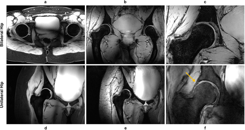
Figure 4.
Examples of hip images acquired with different contrasts. For the bilateral hip, 2D axial GRE (a), 3D coronal MEDIC (b), and a zoomed version of the MEDIC acquisition (c) are shown. For the unilateral hip, 2D coronal GRE (d), 3D coronal MEDIC (e), and PD weighted TSE (f) images are shown with the latter showing the expected contrast between the labrum and cartilage (yellow arrow). (a), (d), (f) were acquired with lipid suppression and demonstrated excellent performance and contrast.

