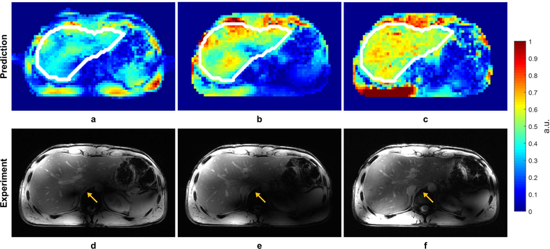
Figure 7.
Predicted B1+ profiles (a, b, c) and acquired low-flip angle GRE images (d, e, f) in the liver with different RF management strategies: static phase-only RF shimming with a homogeneity cost function (a, d); single-spoke pTx pulse (b, e); two-spoke pTx pulse (e, f). The acquired images matched well with prediction in terms of predicted B1+ profiles. The two-spoke pTx pulse effectively mitigated the B1+ inhomogeneity in the liver as demonstrated by improved consistency in contrast throughout the organ (yellow arrows). Note: the experimental data is weighted by the receive profile of the 10-channel dipole array.

