Figure 8.
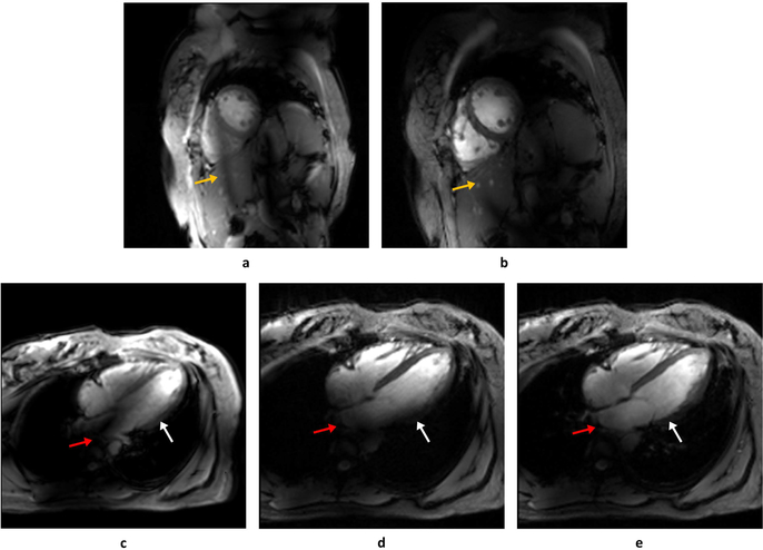
A single frame during diastole of the cardiac CINE acquisitions in the short-axis (a, b) and four-chamber (c, d, e) views. Images acquired before phase-only RF shimming (a, c) are contrasted with those with phase-only static RF shims (b, d). In the 4-chamber view a single-spoke pTx pulse was also implemented (e). In the short-axis view, phase-only shimming improved the myocardium-blood pool contrast and shifted a band of low B1+ outside of the heart (yellow arrows in a, b). However, in the four-chamber view, the phase-only shimming yielded persistent B1+ inhomogeneities in the base of the left ventricle (white arrow) and atrium (red arrow) which were addressed with a single-spoke pTx pulse (e).

