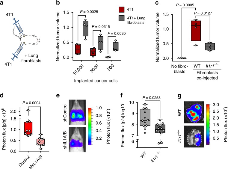
Fig. 6. IL-1 signaling promotes tumor initiation and metastatic colonization of the lung.
a Diagram of tumor initiation experiment where 4T1 mouse mammary tumor cells were subcutaneously implanted alone or co-implanted with primary mouse lung fibroblasts into the flanks BALB/c mice. b Relative tumor sizes in NSG mice after subcutaneous implantation of 4T1 cells in limiting dilutions, alone or in combination with lung fibroblasts; n = 4 mice per group. Tumor sizes were normalized to average tumor size established by 10,000 4T1 cells co-injected with lung fibroblasts. P values were calculated by unpaired one-tailed t-tests. c Relative tumor sizes in NSG mice after subcutaneous injection of 50 MDA-LM2 breast cancer cells alone or in combination with lung fibroblasts obtained from WT or Il1r1−/− C57BL/6 mice; n = 4 mice per group. Tumor sizes were normalized to average tumor size established by MDA-LM2 cancer cells co-injected with WT lung fibroblasts. P values were calculated by ordinary one-way ANOVA with Tukey’s multiple comparisons test. d Lung colonization in mice injected intravenously with control or shIL1A/B-transduced MDA-LM2 breast cancer cells as determined by bioluminescence. P value was calculated by unpaired one-tailed t-test; n = 10 mice per group. e Representative luminescence images from each group in d. f Lung colonization in WT or age-matched Il1r1−/− C57BL/6 mice injected intravenously with E0771 mammary cancer cells as determined by ex vivo lung bioluminescence 24 days post injection. P value was calculated by unpaired one-tailed t-test; n = 12 mice per group from three independent experiments. b–d, f Boxes depict median with upper and lower quartiles, whiskers indicate minimum and maximum values, and data points show biological replicates. g Representative ex vivo bioluminescence images from each group in f.

