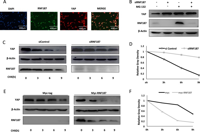
Fig. 5. RNF187 promotes YAP protein degradation.
a Intracellular localization analysis of RNF187 and YAP by immunofluorescence assay. BT549 cells were cultured in normal medium before fixation. Intracellular localization of YAP (red) and RNF187 (green) were shown. Nuclei (blue) were stained with 4′,6-diamidino-2-phenylindole (DAPI). b In the presence of the proteasome inhibitor MG132, the degradation effect of RNF187 on YAP did not further increase YAP protein levels. BT549 cells were transfected with siRNF187 or siControl. After 24 h, cells were treated with 10 µM MG132/vehicle for 6 h. Cell lysates were prepared for Western blot analysis. The results are representative for three independent experiments. c, d RNF187 depletion increased YAP half-life in BT549 cells. BT549 cells were transfected with 50 µM siControl or siRNF187. After 24 h, cells were treated with 100 µM cycloheximide/vehicle for indicated times. Cell lysates were prepared for Western blot analysis. The results are representative for three independent experiments. The YAP relative density was measured by Image J software. e, f RNF187 decreased YAP half-life in HEK293 cells. HEK293 cells were transfected with 0.5 µg Myc-RNF187 or Myc plasmids. After 24 h, cells were treated with 100 µM cycloheximide/vehicle for indicated times. Cell lysates were prepared for Western blot analysis. The results are representative for three independent experiments. The YAP relative density was measured by Image J software.

