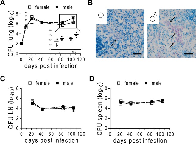
Figure 3.
Bacterial burden in HN878 infected male and female C57BL/6 mice. Females and males were infected via aerosol with a low dose of HN878. CFU were determined in homogenates of lung (A), lymph node (LN; C), and spleen (D) at different time points. (B) PFA-fixed, paraffin-embedded lung tissue sections from day 105 post infection were subjected to ZN staining for detection of HN878 (arrows). Representative micrographs from one mouse out of five mice per group are shown. Bar = 50 µm. (A,C and D) Data is presented as mean ± SD (n = 5). Statistical analysis was performed by Student’s t-test. ***p ≤ 0.001.

