Figure 1.
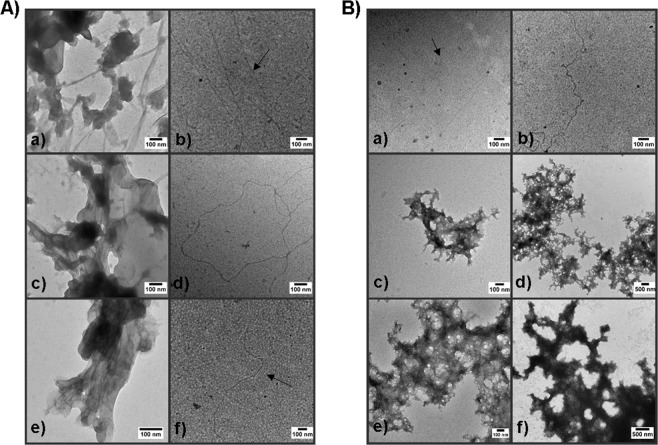
Transmission electron microscopy (TEM) of amyloid mats and purified fibers (A) TEM images of S. mutans amyloid before and after removal of residual monomer. a, c and e) Induced amyloid produced from purified C123, AgA, and Smu_63c, respectively. b, d and f). Amyloid material produced from C123, AgA, and Smu_63c following proteinase K digestion. (B) TEM of purified C123 amyloid fibers incubated without stirring with and without added monomers. a, c, and e) Purified C123 fibers incubated for 2 weeks alone (a) or with added C123 monomer at final concentrations of 1% (c) or 10% (e). e, d, and f) Purified C123 fibers incubated for 4 weeks alone (b) or with added C123 monomer at final concentrations of 1% (d) or 10% (f).

