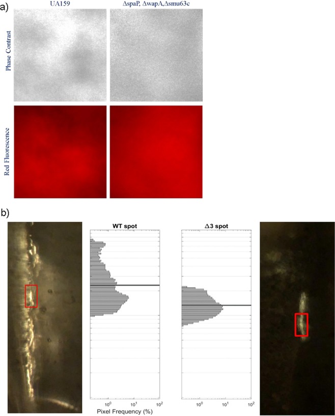
Figure 6.
Assessment of amyloid formation during S. mutans biofilm growth in a microfluidics system. (a) Phase contrast and red fluorescence images following staining of 72 h biofilms of S. mutans wild-type and a triple mutant (Δ3) lacking P1 (encoded by spaP), WapA, and Smu_63c with CDy11. Similar patterns of dye binding to both strains suggests CDy11 reactivity with non-amyloid material. (b) CR-induced birefringence of 72 h biofilms of S. mutans wild-type (left) and the Δ3 triple mutant (right). Insets indicate pixel frequency of the brightest visual locations (red boxes) in each image. In contrast to staining with CDy11, biofilms of the WT and mutant strains could be differentiated on the basis of CR-induced birefringence.

