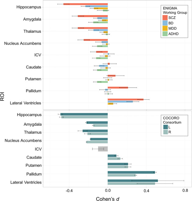
Fig. 5. Subcortical abnormalities in schizophrenia, bipolar disorder, major depressive disorder, and ADHD.

a ENIGMA’s publications of the three largest neuroimaging papers on schizophrenia (SCZ), bipolar disorder (BD), and major depressive disorder (MDD), suggested widespread cross-disorder differences in effects (van Erp et al.54, Hibar et al.68). By processing 21,199 people’s brain MRI scans consistently, we found greater brain structural abnormalities in SCZ and BD versus MDD, and a very different pattern in attention-deficit/hyperactivity disorder (ADHD; Hoogman et al.7). Subcortically, all three disorders involve hippocampal volume deficits—greatest in SCZ, least in MDD, and intermediate in BD. As a slightly simplified ‘rule of thumb’, the hippocampus, ventricles, thalamus, amygdala and nucleus accumbens show volume reductions in MDD that are around half the magnitude of those seen in BD, which in turn are about half the magnitude of those seen in SCZ. The basal ganglia are an exception to this rule—perhaps because some antipsychotic treatments have hypertrophic effects on the basal ganglia, leading to volume excesses in medicated patients. In ADHD, however, the amygdala, caudate and putamen, and nucleus accumbens all show deficits, as does ICV (ventricular data is not included here for ADHD, as it was not measured in the ADHD study). A web portal, the ENIGMA Viewer, provides access to these summary statistics from ENIGMA’s published studies of psychiatric and neurological disorders (http://enigma-viewer.org/About_the_projects.html). b Independent work by the Japanese Consortium, COCORO, found a very similar set of effect sizes for group differences in subcortical volumes between schizophrenia patients and matched controls.
