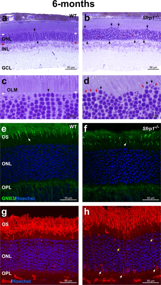
Figure 3.

Morphological alterations of Sfrp1−/− retinas become more evident with age. (a–d) Semi-thin plastic frontal sections from 6 months-old wt and Sfrp1−/− retinas stained with toluidine blue. Black arrows in a,c indicate an organized column of photoreceptors, whereas white and red arrows indicate the outer and inner limit of the ONL. In b and d black and red arrows point to misplaced photoreceptor nuclei and discontinuous OLM respectively. (e–h) Frontal cryostat sections from 6 months-old wt and Sfrp1−/− retinas immunostained with anti GNB3 (cones) or Rho (rods) antibodies and counterstained with Hoechst. White arrows in e,f point to the OS. Yellow and white arrows in h indicate accumulation of Rho in rod processes and cell bodies respectively. Abbreviations. GCL: ganglion cell layer; INL: inner nuclear layer; on: optic nerve; ONL: outer nuclear layer.
